Ectoparasites
 4 December 2018
 12 minute read
 By:
By: Ectoparasites are parasites that live outside the body and their importance varies greatly among regions because of differences in climate and system types used to raise pigs. These parasites feed and reproduce on the host animal at the pig’s expense and especially damaging to young and growing pigs. The extra stress associated with a parasite infestation can cause slow weight gains, adding up to more feed required per kilogram of gain than with an uninfected pig. External parasites produce a range of clinical signs in pigs including rubbing, scratching, and skin lesions. Some parasites also cause significant economic effects due to reduced growth rate, reduced feed efficiency, and loss of carcass value at slaughter.
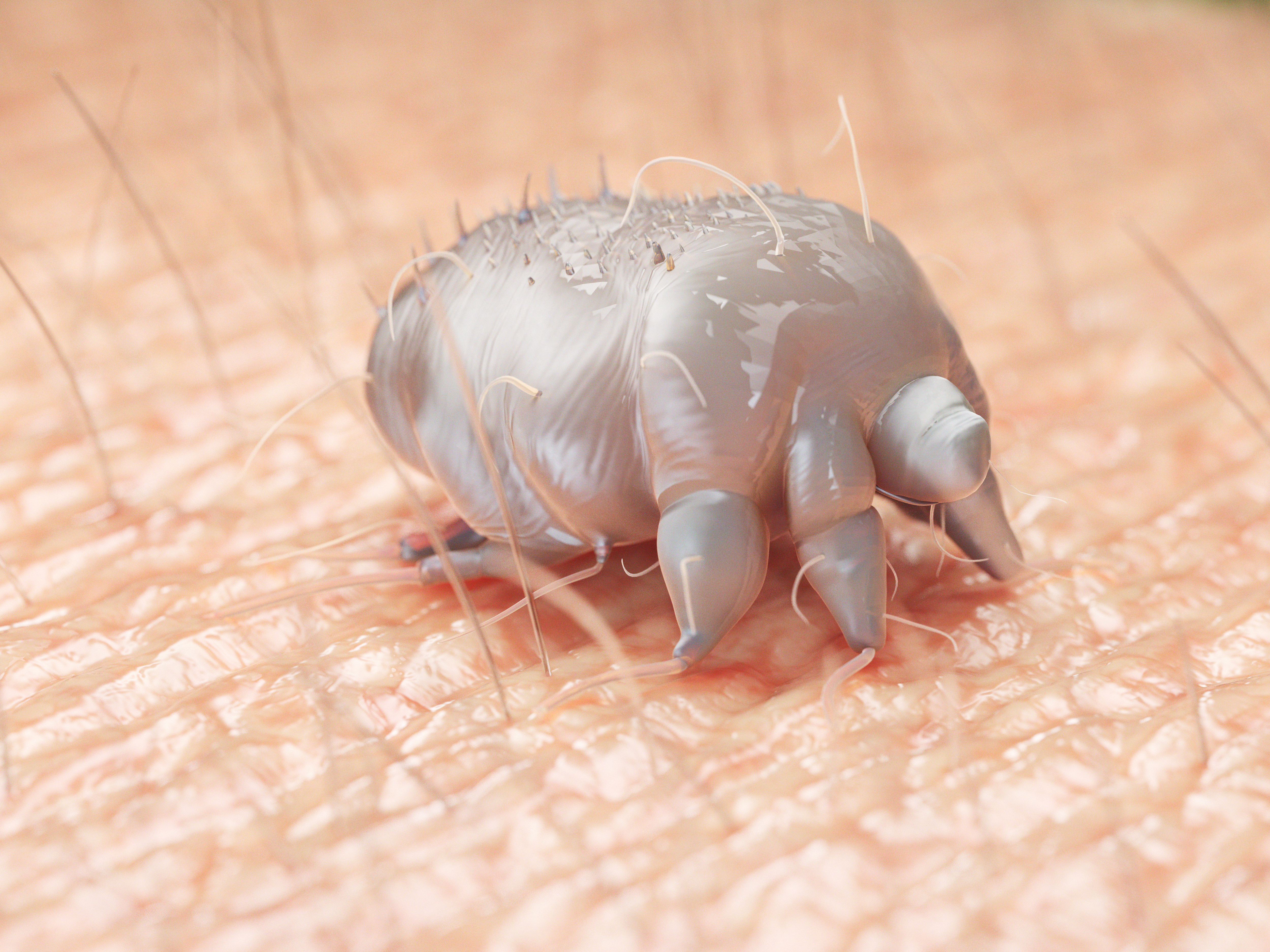
Pig mange mite (Sarcoptes scabiei var suis)
Mange mite is considered the most important external parasite of pigs globally. The sarcoptes mite is a small, greyish-white, circular parasite about 0.5mm in length and just visible to the naked eye when placed on a dark background.
Life Cycle
The life cycle stages are egg → larva → nymph → adult, all of which occur on the pig host. The adult female mite burrows tunnels to beneath the skin surface, where she lays up to 60 eggs over a period of 30 days. The eggs hatch in about five days, and the larvae may remain in the parent tunnel or start new tunnels. The cycle from egg to fertilised female takes 10 to 15 days and many generations of mite may be on one pig.
Clinical disease
Two clinical forms of the disease are recognised:
- A hyperkeratotic form that most commonly affects older, multiparous sows and form encrustations in the ears of sows and considered the main reservoir of mites within a herd, infecting piglets during nursing. Boars may become chronic carriers.
- A pruritic (itchy) or hypersensitive form that primarily affects growing pigs, particularly when closely housed in groups.
Acute disease
Common signs include ear-shaking and severe rubbing of the skin against the sides of the pen. Approximately three to eight weeks after the initial infection, the skin becomes sensitised to the mite protein and a severe allergy may develop in susceptible individuals with very small red pimples covering the complete skin. The intense irritation may cause intensive rubbing and bleeding occurs.
Chronic disease
After the acute phase, thick encrustations, full of mites, develop on the ear, along the sides of the neck, the elbows, the front parts of the hocks, and along the top of the neck.
Diagnosis
The diagnosis of sarcoptic mange is confirmed by proving the presence of the mite in the herd. The best method is to use a torch to examine the internal surface of the ears of breeding animals for encrusted lesions. Using an implement e.g. teaspoon encrusted lesions can be scraped off and placed on a black piece of paper and left undisturbed for 10 minutes. After 10 minutes tip the paper upside down to remove the scabs, any mites present will adhere to the paper and can be seen with the naked eye or with a magnifying glass.
Establishment and maintenance of mange-free pig populations
The mite dies quickly in the environment and under most farm conditions only lasts up to five days typically but up to 4 weeks in moist protected conditions. Mites are sensitive to drying, and if they are exposed to direct sunlight or dry surroundings, they will not survive for more than 24 to 48 hours. This is an important factor in control. When a herd is free from mange, it is one of the easiest diseases to keep out because it can only be introduced by carrier pigs. However, once it is introduced, it usually becomes endemic unless control measures are taken. Exposure to environments where the mites are still alive will result in an infection for naïve pigs in as little as 24 hours.
The establishment and maintenance of mange-free herds is expedited by three important facts:
- Piglets are born free of mites
- Mites are highly host-specific and do not survive long away from their host, and
- Modern treatments are very effective.
Control
Mange control involves identification of animals with chronic mange so that they can receive systematic and regular treatment to protect the younger animals in the herd. All control programmes must target the breeding herd. All animals with extensive hyperkeratotic lesions in the ears and over the body should be culled and the remainder of the sows treated simultaneously or alternatively in segregated groups prior to farrowing.
Contaminated bedding should be removed and the environment sprayed with insecticide.
- Treat all pigs regularly to prevent a build-up of numbers.
- Treat boars every three months.
- Always treat animals twice, 10 to 15 days apart.
- Leave pens empty for five days after infected pigs move out, perform cleaning, disinfection and drying procedures followed by spraying with an appropriate insecticide.
Mange-free herds can be established by depopulation and repopulation from mange-free stock, by segregated rearing of treated pigs or by eradication using ‘avermectin’ type products and other products registered for the purpose.
Implementing bio-security measures especially on isolating and treating incoming stock and sourcing stock from a minimal number of herds are usually adequate to prevent re-introduction of the parasite.
Demodectic mange (Demodex phylloides)
This mite is considered relatively unimportant in pigs. It lives in hair follicles. The response to treatment is poor but the mite is sensitive to those acaricides used for sarcoptic mange control. Severely affected animals should be culled from the herd.
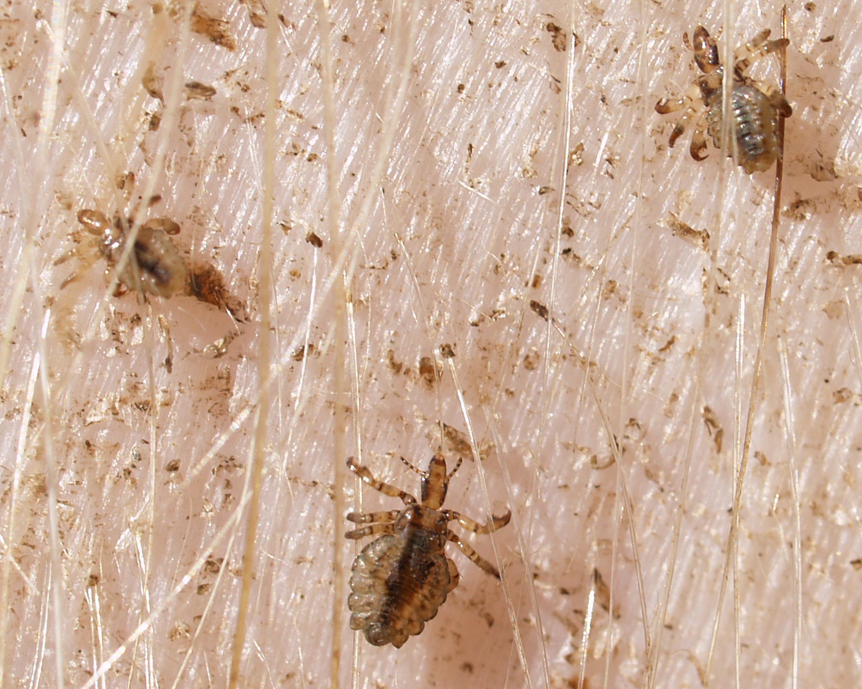
Pig lice
Haematopinus suis is the louse that affects pigs. It has piercing and sucking mouthparts and is greyish-brown in colour with brown to black markings, although may take on a blueish appearance after feeding. The females are about 6mm long and the males slightly smaller and so are easily seen with the naked eye, especially on a white breed.
The pig louse is host-specific and cannot survive for more than two to three days away from pigs with the lifecycle completed from egg to adult in 23 to 30 days. Lice are found on all parts of the body, but particularly in the folds of the skin around the neck, jowl, flanks and on the inside of the legs - where it is slightly warmer and protected. Lice can often be found inside the ears seeming to form nests. Transmission is primarily by direct contact although naïve pigs being put in a recently vacated infected pen can become infected.
Lice are often blamed for damage caused by mange because both conditions cause irritation and rubbing. Lice should be relatively uncommon in herds today as treatments are readily available. Heavy infestations result in anaemia in young pigs and may affect growth rate and feed efficiency. The heaviest infestations of pig lice usually occur in winter.
Life cycle
The adult female attaches 2-4 eggs per day to hair shafts with a cement like substance and may lay as many as 90 eggs over a 25-day period. These yellow eggs can be seen easily on dark haired pigs. The young lice (nymphs) emerge from the eggs in 10 to 21 days, maturing to adults by 30 days, depending on environmental conditions. The nymphs go through three developmental stages and feed on blood in all three stages before reaching the adult stage. The average lifespan for male and female lice is about 25 days.
Treatment and control
Treatment and control of lice is easily achieved because the mites live on the skin surface and can survive only a few days away from their host. Treatments may be applied to the pig in the form of sprays, pour-ons, injections, and as in-feed medications. Two doses 10-14 days apart will eliminate lice. All treatments are ineffective against eggs hence the need to treat twice.
Control and eradication strategies listed for sarcoptic mange apply equally for lice. These include special attention to the ears, treatment of the boars, multiple treatment of sows prior to farrowing, segregation of clean and untreated animals if the whole herd is not treated at one time, and treatment of all introduced animals.
Ticks
Ticks infest many species of mammals and birds and are not host-specific, due to the pigs skin they are rarely an issue and only occasionally seen in outdoor herds.
Ticks are easily seen by gross visual examination and their size and appearance dependent upon if they have recently fed or not. They can be found on any part of the body, but they are more often seen around the softer skin of the ears, neck and flanks.
The treatment and control of ticks in pigs are rarely required. If only a few ticks are present, these can be removed manually with a tick removal tool and the pigs confined away from infested pasture. Treatments licensed for lice control are usually effective and there are no products licensed specifically for tick treatment.
Flies
Flies can be a concern in pig production for disease transmission and welfare reasons as such fly populations are often used as a measure of hygiene by assurance schemes.
Some flies annoy animals by their vicious bite, while others act as a vehicle for transmission of infectious disease.
Flies make contact with faeces, skin, and mucus and milk discharges of the pig. When they reach a high enough level they can both irritate the pigs by biting (depending upon fly species) and become major transmitters of disease causing organisms such as pathogenic strains of E. coli, B. hyodysenteriae, salmonella, streptococci and rotavirus.
Major outbreaks of greasy pig disease and coccidiosis can be exacerbated by high fly populations and when sows have mastitis, flies are attracted to the udder and skin surfaces in great numbers and they can be responsible for enhancing severe outbreaks.
Fly control in all piggeries must be continuous in summer months and detailed in farm health plans with the aim to prevent flies from breeding and to destroy adult flies. Breeding of flies can be prevented by the regular removal of dung and using baited fly traps and insecticides.
Mosquitoes
In endemic areas mosquitos may attack livestock causing discomfort at best and severe irritation at worst. Lesions can appear on several or all of the pigs in the form of raised oedematous weals on the legs and abdomen. In severe cases affected carcasses of pigs must be skinned at slaughter. Mosquitoes are important vectors in the transmission of Japanese encephalitis virus and act as mechanical vectors in the transmission of Eperythrozoon suis. Mosquito bites can irritate nursing sows resulting in increased overlays and piglet deaths.
There are several control measures that can be implemented to decrease the number of mosquitoes. Where there is a disease outbreak fogging may be considered as an option in order to kill the infected adult mosquito population and local councils may use larvicides to prevent human infection. Where possible, the breeding ground of the mosquitoes should be identified and the larvae destroyed by either draining water reservoirs or covering the surface with environmentally safe oil.

Emily Houghton
Emily Houghton is a Zoology graduate from Cardiff University and was the editor of The Pig Site from October 2017 to May 2020. Emily has worked in livestock husbandry, and has written, conducted and assisted with research projects regarding the synthesis of welfare and productivity of free-range food species.